


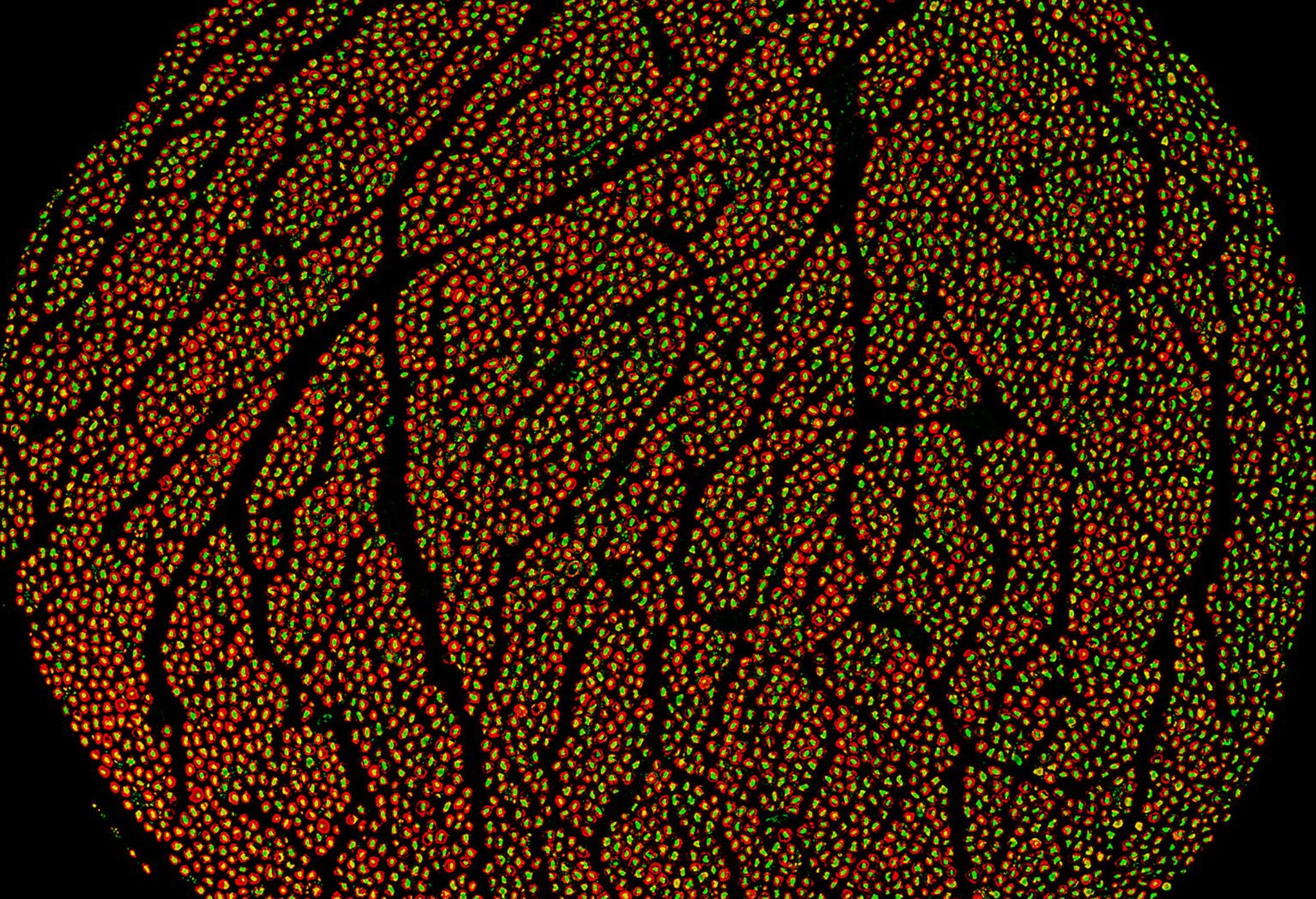
Congratulations to Feinstein Institutes for Medical Research, which have released a genuine advance on the received anatomy of the human vagus nerve!
Zanos, S., Jayaprakash, N., Khaled, Q., Nasrallah, Z., Barbe, M., Chen, F. L., miller, larry, Zanos, T., Levy, T. J., Vardhan, A., Cang, J., Toth, V., Coppa, K., Ben-Shalom, N., Song, W., Carpentiere, N., Kanavos, T., Birbas, E., Bahadir, S., & Saleknezhad, P. (2025). Human vagus nerve anatomical reconstruction using microCT immunohistochemistry and ultrasound - f011 [Dataset]. In SPARC REVA FEINSTEIN (Version 1). SPARC Portal. https://doi.org/10.26275/BK6F-QSKP
MicroCT imaging at nine-micron resolution, performed continuously from the cervical to the abdominal vagus, resolves fascicular architecture — how fibers are bundled, how those bundles subdivide and reorganize along the nerve’s length — at a scale no unaided dissection has ever approached. Cross-registered against ultrasound and immunohistochemistry, the dataset does something more interesting than simply seeing further into a familiar structure. Its IHC panel — neurofilament, myelin basic protein, tyrosine hydroxylase, choline acetyltransferase — stains for neurochemical identity rather than relying on visual salience alone, asking of the tissue not merely “is this fiber large enough to have survived and be seen” but “is this fiber sympathetic or parasympathetic in character.” This is a genuinely different question than gross dissection has ever been equipped to ask, and it is the closest a necrotic-tissue protocol has come to addressing, rather than merely restating, the trunk-line problem.
(What is the TRUNK LINE PROBLEM? See below at the end of this essay.)
Yet it is precisely because this achievement is substantive that the entire project’s framing deserves particular scrutiny. The Feinstein Institutes’ own announcement describes the release as making public the “world’s first comprehensive human vagus nerve maps, solidifying decades of leadership in bioelectronic medicine.” Both halves of that sentence greatly overreach. “Comprehensive” is a claim of completeness the dataset’s own curators do not make for it — their notes describe a single female cadaver, a preliminary release intended to test publishing and processing pipelines, with more data and protocols promised for later. A single-subject, structurally partial dataset, honestly and usefully partial, is not comprehensive by any ordinary meaning of the word despite being a substantial improvement in resolution over what preceded it. And “solidifying decades of leadership in bioelectronic medicine” does something the anatomy itself cannot do on its own terms — it borrows the neutrality of an atlas to underwrite a claim of institutional and commercial standing in a therapeutic field, a claim that belongs to the register of marketing, not to the register of anatomical description. The gap between what the curator’s notes disclose and what the press release announces is the tell: evidence that the promotional register and the scientific register are not equivalent.
Yet the failure runs deeper than the rhetorical framing, and it runs in two independent senses.
The first sense is structural: the fine fiber network is not visualized here, at any resolution this protocol reaches. MicroCT’s nine-micron voxel size is an order of magnitude too coarse to resolve individual unmyelinated fibers, which run from the sub-micron to low single-micron range; the one technique that could in principle catch them, immunohistochemistry, is deployed at five sampling levels per half-centimeter subsegment — a sparse set of static cross-sections, not a continuous account of the terminal network along the nerve’s length or within the organs it innervates. What results, however much finer than anything that preceded it, is a better map of the trunk lines and a genuinely improved map of the interior of the trunk — how the grain runs, how the major and minor boughs are internally organized. It is not a map of the branches, and it is not a map of the leaves, and by the instruments this protocol uses, it cannot be. In this first sense the dataset will never be structurally complete because the tissue category that would have to be visualized to complete it does not survive long enough, in a form these instruments can resolve, to be imaged at all. This is therefore not a complete mapping of the vagus, nor can it ever be irrespective of how large the data set becomes, or how many cadavers are thusly dissected. It is a map of the neurons of the vagus that survive cell death, which is to say a map devoid of the fine fiber network.
The second sense is not structural, and would remain true even if the first problem were somehow solved — even if some future instrument resolved every fiber down to its terminal ending. It is that the entire premise motivating this kind of anatomical refinement — that finer-grained mapping of fascicular structure enables more precisely targeted vagal stimulation, and that more precise targeting should be expected to yield proportionally better therapeutic outcomes — is trunk-line logic applied at higher resolution and still partakes of a category error.
Autonomics holds that a fiber’s behavioral and physiological meaning is not fixed to its anatomical address. It is a function of the pervading neurochemistry the fiber is embedded in at the moment of activation — the dynamic, state-dependent neurohormonal and neurotransmitter environment that determines what a given circuit is doing, and is capable of doing, at that moment. Two identical fascicles, in two different autonomic states, do not mean the same thing when stimulated. If this is correct, then knowing which fascicle a fiber runs in, however precisely, answers a structural question that the therapeutic question was never actually asking. Precision targeting of trunk-line structure, absent any account of the context the structure is operating within, should be expected to produce diminishing returns almost by construction — not because the engineering is poor, but because the variable being refined is not the variable that determines the outcome.
This is not a speculative extension of the methodological critique above; it follows directly from it. The one modality in the protocol capable in principle of imaging a living, contextualized nerve — ultrasound — was performed on the same fixed cadaver as everything else, foreclosing the possibility before the imaging began.
What the neurochemical stains register, however precisely, is fixed molecular identity in dead tissue: what a fiber was built to carry, not what state it was carrying it in. That is the specific mechanism by which a comprehensive structural map, however well executed, cannot become a map of function.
The dataset sharpens the trunk and clarifies its interior structure past anything available before it. It does not, and by its own methodology cannot, reach the branches and the leaves it never visualized, and even where it succeeds in resolving structure, it cannot supply the state-dependent context that gives that structure its meaning. Two absences, not one — and no increase in imaging resolution addresses either of them.
WHAT IS THE TRUNK-LINE PROBLEM?
