In vivo imaging of the living Autonomic Nervous System

I didn’t cut anybody open, ok? I’m not vivisecting anyone.
Let’s just start there. I have been on and on for a number of years about inaccurate neuro-anatomical depictions of the Vagus, and why they matter. I was messing around last week with some technique in Photoshop, trying to find a way to make an image look like a woodblock print, and had the inspiration to start playing with this as an imaging tool to help people grasp, more effectively, their living neurology.
If you go out looking for a decent illustration of the Vagus, you are going to find something about like this. Pay attention in particular to the circled area at bottom, which is the subdiagphragmatic Vagus. This is what we refer to as the Grounding System, what Porges calls Dorsal Vagal. It is largely unmyelinated.
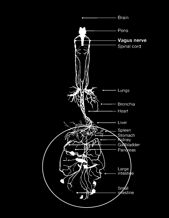
You would be left with the impression, hereby, that the neural conduits into the guts are a rather loose affair. A few wires in a loose basket.
This would be false.
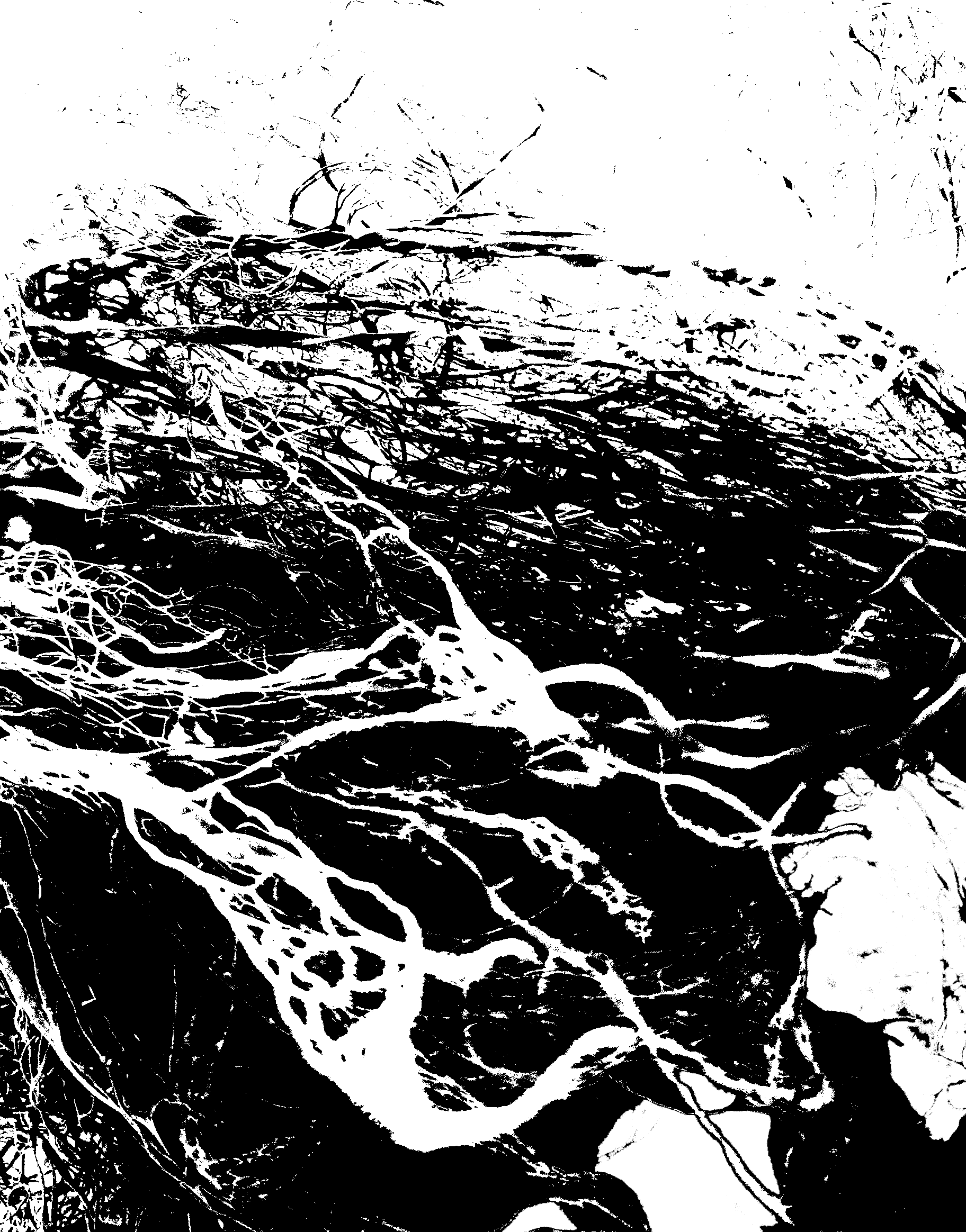
If you zoomed in and looked at the texture of the actual living autonomic neurology in the guts, it would look more like this: